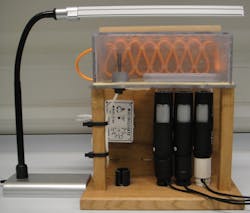
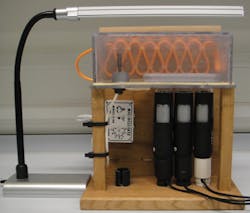
Light microscopy systems for measuring cell motility can cost hundreds of thousands of dollars. But a PhD student at Brunel University London's College of Health and Life Sciences (Uxbridge, England) built his own inverted microscope for an estimated $250 by adapting three low-cost USB microscopes he purchased online.
In learning about cell motility tracking, Adam Lynch, a student in the College's Institute for the Environment, learned how a snail's immune system responds to chemical pollutants present in water, which might influence transmission of Schistosome parasites to humans (the parasites can induce chronic infection of the urinary tract or intestines in humans). Lynch and his collaborators needed more than one inverted microscope to run multiple tests, but wanted to avoid the high costs associated with having more than one system.
Lynch realized that the three Veho VMS-004D 400x USB microscopes (each with 1.3 Mpixel CMOS image sensors) he had purchased could be clamped upside down on a table (for stability) to produce the same images that a far more expensive inverted microscope can. His system, which he calls the Low-Cost Motility Tracking System (LOCOMOTIS), involved creating a 3D model using an open-source software program, followed by its frame and stage construction. For illumination, an external LED strip light was used due to its low heat emission and intensity, which helps reduce stress to the cells.1
Lynch points out that getting the right angle of lighting enabled the LOCOMOTIS system's success. When he turned off the USB microscopes' onboard LED illumination and instead used external illumination, he found that he could see the cells quite clearly—the system allowed him to observe cells that measured about 50 µm long.
The next step for Lynch and his collaborators is to determine additional utilities for LOCOMOTIS, and to lower its cost even further.
1. A. E. Lynch, J. Triajianto, and E. Routledge, PLoS One, 9, 8, e103547 (2014).


