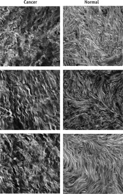
Second-harmonic-generation (SHG) microscopy is one of the nonlinear microscopic imaging techniques widely used in life sciences research to acquire three-dimensional images that provide structural and functional mapping information. Because these images allow microscopic pathology (examination and interrogation) of tissue samples from biopsy and even in situ in patients, nonlinear microscopy is finding its way into preclinical and ultimately clinical applications. This translational trend is supported by a new type of ultrafast fiber laser offering the requisite long (1055 nm) wavelength in the necessary compact, rugged, maintenance-free format.
Ultrafast 3D MPE microscopy
Because the first demonstration of nonlinear microscopy using ultrafast lasers involved multiphoton excitation (MPE) of fluorescence, the various related techniques are often collectively referred to by the term MPE. These MPE methods have revolutionized optical microscopy for life sciences because they provide inherent three-dimensional resolution in living specimens with little or no damage. Due to the two- (or three-) photon absorption process, the beam intensity drives the nonlinear effect only right at the beam waist. This eliminates the need to use a pinhole aperture to spatially filter out background light from the signal, as in one-photon confocal microscopy. A three-dimensional image is built-up by rastering the laser beam on the image plane (x-y) and moving the sample, or the objective, in the z direction.
Common examples of nonlinear imaging include two- and three-photon excitation of fluorophores (MPE), coherent anti-Stokes Raman scattering (CARS), second-harmonic generation (SHG), and third-harmonic generation (THG). In addition, there is a growing range of practical variations on the way these techniques are implemented, such as spatial light modulators or multibeam methods aimed at reducing the time it takes to acquire an entire image plane (sectional image) or image cube.
Beyond three-dimensional discrimination, there are additional advantages of nonlinear microscopy that make it ideal for tissue imaging. First, the low probability of these nonlinear effects means that the sample absorbs only a tiny fraction of the incident laser light along the beam path, while the rest is scattered in many directions. Contrast this with confocal microscopy, where most of the light is absorbed on the beam path. This low absorption greatly limits any photodamage that could compromise, or even kill, the sample.
And, whereas fixed tissue can be arbitrarily sectioned as needed, imaging live tissue usually requires imaging deep inside the tissue. This is certainly the case for any emerging uses of nonlinear microscopy for in situ biopsy of human tissues. Light scatter is the main limiter on the depth at which any imaging can be performed. In most materials, scattering is inversely related to wavelength, with longer wavelengths enabling deeper imaging. This favors nonlinear techniques that use wavelengths two or three times longer than the visible light used in conventional or confocal microscopy.
Why SHG?
Most two- and three-photon excitation microscopy use fluorescent stains and dyes. More recently, genetically encoded fluorescent probes and indicators have enabled a range of new techniques that are helping to answer several questions in biology.
Because dyes or genetic expressions cannot be used with human subjects, they are outside the reach of real-time, in situ biopsy. It is generally not practical or safe to inject human subjects with high concentrations of most of these fluorescent stains and indicators, and of course it is even more impractical or unethical to use genetic modifications! Thus, the only MPE methods that are good candidates for use on humans or clinical samples are those that rely only on endogenous fluorescent materials present in tissue. For example, harmonic generation methods like SHG and THG can be used to image NADH, while CARS/SRS techniques are excellent for imaging lipids.
Simplicity and a pathway to lower-cost automated testing are prerequisites for any method contemplated for transition to future clinical use. This somewhat limits CARS, which is arguably the most complex of all MPE techniques, as it requires two different ultrafast beams at different wavelengths that are optically aligned and temporally synchronized. In addition, one of the CARS wavelengths is typically around 800 nm, limiting tissue penetration and usable power before onset of damage. Similarly, endogenous fluorophores like NADH typically require excitation at 710–800 nm. This leaves SHG and THG as potential candidates for preclinical and clinical purposes.
As their names indicate, in SHG and THG, a fraction of incident laser light is coherently converted to the second- or third-harmonic wavelength when certain optical conditions are met. In theory, THG can occur at most interfaces between different refractive indices, making it useful for imaging cell structure boundaries or lipid water interfaces. However, to generate a signal in the visible spectrum, THG needs excitation at wavelengths hard to access with a simple laser oscillator (1.2–1.5 μm). Thus, preclinical use is bound to be limited until there is an economical path to producing these wavelengths.
SHG occurs whenever a laser with high peak power is focused into non-centrosymmetric structures (see photo at top of this page). Some of these occur naturally, with the most common example being collagen found in muscle and many other tissues. Just as important, SHG is practically wavelength-independent, thus enabling the use of newly available, simple ultrafast laser sources at convenient wavelengths. Because of these advantages, SHG has become the leading candidate for preclinical studies and probably represents about 80% of the current research in preclinical in vivo nonlinear imaging.
New, simple lasers at 1055 nm for brighter images
As already noted, the use of longer wavelengths for any type of nonlinear imaging enables deeper penetration and lower damage. For in vivo SHG, penetration of a few hundred microns is highly desirable, so a longer wavelength is preferred. On the other hand, depending on the actual tissue structure, the intensity of the SHG generated or scattered backward may decrease with increasing wavelength. No definitive study has yet determined the best wavelength for human tissue, but several studies at wavelengths longer than 1000 nm showed these wavelengths to be a good match for SHG.
Titanium:sapphire (Ti:sapphire) lasers are workhorse ultrafast lasers that dominate the majority of nonlinear microscopy applications. But while a few Ti:sapphire lasers can reach 1080 nm, fundamental limitations of the gain material mean that relatively low power levels are available beyond 1000 nm. The use of a tunable optical parametric oscillator (OPO) to extend this range is extremely useful for research and optimization, but it is overkill for preclinical-type work because of cost and complexity.
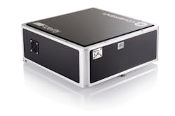
Fortunately, a new generation of high-power ultrafast lasers with fixed output wavelength around 1055 nm has been developed based on diode-pumping of ytterbium-doped fiber. This fiber-based technology is optically simpler than Ti:sapphire lasers and can be economically packaged in a compact and extremely rugged, zero-maintenance, sealed laser head. An example of this next-generation ultrafast fiber laser is the new Fidelity from Coherent (see Fig. 1).A key goal for any potential medical laser tool is to provide maximum effectiveness with minimum total dosing. For in vivo nonlinear imaging, this means achieving the brightest possible image for a given laser's average power level. In SHG and all these nonlinear techniques, image brightness is proportional to the product of peak power and average power. Higher peak power can be achieved either by increasing the pulse energy and/or decreasing the pulsewidth, as long as sample damage is avoided. Fidelity meets this goal by delivering much shorter pulsewidths (<70 fs) than other ultrafast fiber lasers (which are typically 100–250 fs) at a low-damage wavelength.
Of course, it is the peak power and average power at the sample that actually determines the brightness of the SHG images. The broader spectral bandwidth of a 70 fs pulse means that group velocity dispersion (GVD) in the beam delivery optics will stretch the initial laser pulsewidth. For this reason, Fidelity incorporates a software-controlled pre-compensator to enable the user or system builder to offset downstream GVD and thus minimize the pulsewidth at the sample. Moreover, this feature allows researchers the option to smoothly vary the pulsewidth at the sample, and thereby study how image brightness and any accompanying photodamage scale with pulsewidth.
Maximizing productivity
The newest generation of ultrafast fiber lasers delivers an optimum combination of output characteristics for SHG microscopy, especially when deep tissue imaging is required. Plus, these new sources offer enhanced reliability and ease of use, together with high data acquisition rates, enabled by their combination of very high repetition rate and high average power. Together, this maximizes the productivity of MPE microscopes based on these lasers, and makes this technology accessible to the widest possible audience.
REFERENCE
1. C.-Y. Dong et al., J. Biomed. Opt., 18, 3, 031101 (Apr. 5, 2013); doi:10.1117/1.JBO.18.3.031101.

