Introduction
Optical Coherence Tomography (OCT) has established itself as a groundbreaking technique, heralding a new era in non-destructive three-dimensional (3D) imaging of various materials and tissues. Rooted in the principles of Fourier Transform and image generation, OCT has found applications across a spectrum of fields, including research, medical diagnostics, guided surgery, industrial processing, and non-destructive testing. This article endeavors to offer a comprehensive insight into OCT’s operational mechanisms, its significance in diverse domains, and its profound impact on disciplines such as ophthalmology and neuroscience.
Optical coherence tomography (OCT) represents a non-invasive, high-resolution optical imaging technology that harnesses the power of interference signals to construct cross-sectional images. These images are derived from interactions between a subject under investigation and a reference optic, making OCT a pivotal tool in medical applications where real-time, two-dimensional (2D), and three-dimensional (3D) in-vivo images are essential for visualizing tissue structures.
OCT plays a pivotal role in the enhancement of diagnosis and management of ophthalmic conditions such as Age-related Macular Degeneration (AMD), a retinal disease that results in blurred vision, and diabetic retinopathy. OCT quantitatively characterizes structural and appearance changes in retinal tissue, enabling more effective therapies. By tracking retinal thickness and biomarkers, OCT offers insights into disease progression. Furthermore, OCT can be adapted for cardiology, allowing the early diagnosis of conditions that may lead to heart attacks. Atherosclerosis, a primary cause of heart attacks, is marked by ruptured fatty plaques and calcium buildup within arterial walls, obstructing blood flow. OCT is instrumental in the detection of vulnerable plaques prior to rupture by providing an image resolution of 5-7 µm to ascertain plaque size, shape, and location. In both ophthalmology and cardiology, early and precise diagnosis transforms treatments and aids in unraveling the cellular progression and pathways of numerous incurable diseases.
Working Principle of OCT
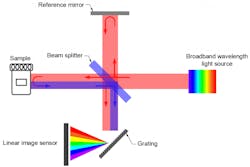
At its essence, OCT functions as an interferometric imaging technique, relying on the interference of light waves. It leverages low-coherence interferometry, using a broadband light source that emits near-infrared light, typically ranging from 800 to 1,300 nm. This light is divided into a sample arm and a reference arm. The sample arm directs light toward the subject or tissue of interest, while the reference arm houses a reference mirror. The light reflected from both the sample and reference arms is combined, and the interference between the two beams is precisely measured.
OCT’s power in medical imaging stems from its ability to employ light for capturing high-resolution, three-dimensional images by detecting optical scatter within biological tissue. While there exists a trade-off between depth penetration and resolution, OCT often collaborates with other technologies to ensure accuracy and multifaceted image acquisition.Fourier Transform and Image Generation
To craft 3D images, OCT harnesses Fourier Transform techniques. The interference pattern formed from the combined light beams is processed using Fourier Transform algorithms to unveil depth information. As the sample arm scans along one axis, a series of A-scans (depth profiles) are collected. These A-scans, when amalgamated, create a two-dimensional (2D) cross-sectional image known as a B-scan. By progressively acquiring multiple B-scans, a comprehensive 3D volumetric image comes into existence.
In conclusion, Optical Coherence Tomography stands as a technological marvel that has revolutionized our capacity to see the unseen and diagnose diseases with unprecedented precision. Its potential applications are limitless, and as technology continues to advance, OCT promises to play an increasingly significant role in shaping the future of medical diagnostics and imaging across various disciplines.
Do not hesitate to contact Shanghai Optics today. We’d be more than happy to discuss your projects and how best they can become a success.

